

Trauma
Question 156 of 180
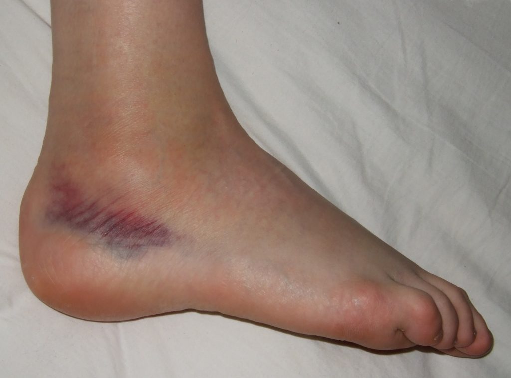
A 19 year old girl walks into the Emergency Department with a painful right ankle. She was out drinking last night, wearing high heels, and remembers falling over. She woke up this morning with a swollen, bruised, painful ankle, which is painful to walk on. On examination you note tenderness inferior to the lateral malleolus. What should the next step in her management be?
Answer:
Management of ankle sprain:- Advise initial rest, elevating the ankle above hip level, and to consider applying ice intermittently during the first 2 days for periods of 10– 15 min.
- Begin to weight- bear as soon as symptoms allow, but elevate at all other times. An elastic support from toes to knee is traditional, but of no proven value (and may be harmful by increased pain without speeding recovery). If used, ensure that it is not worn in bed.
- Advise the patient to gently exercise the ankle in all directions and to use simple analgesia regularly until symptoms improve.
- Most patients with minor sprains can expect full recovery in ~4 weeks. It may be possible to resume sports gradually within 2 weeks, depending on progress.
Ankle Injuries
Trauma
Last Updated: 13th February 2021
Assessment of ankle injuries
- Ankle injuries are amongst the most common problems presenting to the ED.
- Adopt a logical, consistent approach to identify which patients are likely to have a fracture and to avoid unnecessary X-rays in patients with uncomplicated sprains.
- History
- Establish the exact mechanism of injury. Most are inversion injuries (where the sole of the foot turns to face medially as the ankle is plantar flexed) causing damage to structures around the lateral malleolus (most notably, the anterior talofibular ligament). Eversion injuries occur less commonly and damage the structures around the medial malleolus. Hyper-dorsiflexion and plantar flexion injuries occur less frequently.
- The following are relevant in the initial assessment of ankle injuries:
- A fracture is more likely in patients who are unable to weight- bear immediately following the injury.
- A ‘crack’ or ‘snap’ may be heard and is not indicative by itself of a fracture.
- Ice, analgesia, and elevation may influence the appearance of an ankle injury.
- Examination
- Examine from the knee down for tenderness over the:
- Proximal fibula.
- Lateral malleolus and ligaments.
- Medial malleolus and ligaments.
- Navicular.
- Calcaneum.
- Achilles tendon.
- Base of the fifth MT.
- Examine from the knee down for tenderness over the:
- Is an X-ray required?
- Follow the Ottawa ankle rules for adults and X-ray the ankles if patients:
- Were unable to weight- bear for four steps both immediately after the injury and at the time of examination.
- Have tenderness over the posterior surface of the distal 6cm (or tip) of the lateral or medial malleolus.
- Note that tenderness over the navicular, calcaneum, base of the fifth MT, or proximal fibula require specific X- rays to exclude fractures.
- Adopt a lower threshold for X- ray in the very young, the elderly, and in patients who are difficult to assess (e.g. intoxicated).
- Follow the Ottawa ankle rules for adults and X-ray the ankles if patients:
Ankle fractures
- Fractures around the ankle most commonly involve the malleoli— medial, lateral, and what is commonly referred to as the ‘posterior malleolus’ (the posterior part of the distal tibia). The ankle mortise joint allows very little rotation or angulation at the ankle joint, so forced twisting or angulation of the ankle joint causes fractures associated with ligamentous injuries and, in severe cases, disruption of the distal tibiofibular syndesmosis.
- Treatment depends upon a combination of clinical findings and X- ray appearances. Look carefully for talar shift.
- Small avulsion fractures essentially reflect ligament/ joint capsule damage. Treat with rest, elevation, analgesia, and early mobilisation, as for sprains.
- Larger avulsion fractures may require initial immobilisation in a boot ± crutches and orthopaedic follow- up.
- Undisplaced, isolated medial or lateral malleolar fractures are usually stable and do well with conservative measures. Provide analgesia and crutches, and immobilise in a well- padded BKPOP cast. Advise limb elevation, and arrange orthopaedic follow- up. Note that an isolated ‘high’ lateral malleolus fracture may only be apparent on the lateral X- ray and may be associated with deltoid (medial) ligament injury with instability— some require ORIF.
- Displaced fractures of the medial or lateral malleolus require ORIF. Give analgesia and, as appropriate, IV sedation to allow reduction of talar shift. Immobilise the limb in a BKPOP slab, and refer to the orthopaedic team.
- Bimalleolar or trimalleolar fractures are unstable. Having attempted to reduce any significant talar shift (with appropriate sedation), place in a BKPOP, obtain fresh X- rays, and refer to the orthopaedic team.
Ankle dislocation
- Dislocation of the ankle is a true emergency. Treat promptly on diagnosis. Examination shows gross deformity of the ankle, severe stretching of the skin (resulting in fracture blisters, skin necrosis, or even converting the injury to a compound fracture), and often deficits in peripheral pulses or sensation. The ankle can dislocate in the absence of associated fractures, but this is uncommon.
- Prompt closed reduction and immobilisation in POP usually have to precede X- ray (unless available immediately). ‘Prompt treatment’ does not justify reduction without considering analgesia or sedation.
- Reduction technique
- Give Penthrox®, IV analgesia/ sedation, as needed, with full precautions.
- Warn that there may be a brief increase in discomfort as the ankle reduces.
- With the knee flexed and supported, gently grasp the heel with one hand and support the patient’s calf with the other.
- Pull smoothly on the heel— it may be necessary to slightly exaggerate the deformity in order to obtain reduction. Success is indicated by return of normal ankle contours, relief of skin tension, and often dramatic relief of pain.
- Once reduced, re- check pulses and sensation; immobilise in a POP slab, and arrange check X- rays.
- Refer the patient to the orthopaedic team immediately.
Ankle sprains
- The structures most frequently injured in inversion injuries are the lateral joint capsule and the anterior talofibular ligament. Increased injury causes additional damage to the calcaneofibular ligament and posterior talofibular ligament.
- Historically, treatment of sprained ankles has been based upon ‘RICE’ (rest, ice, compression, elevation), but the scientific basis for all the elements of this is distinctly lacking!
- Advise initial rest, elevating the ankle above hip level, and to consider applying ice intermittently during the first 2 days for periods of 10– 15 min.
- Begin to weight- bear as soon as symptoms allow, but elevate at all other times. An elastic support from toes to knee is traditional, but of no proven value (and may be harmful by increased pain without speeding recovery). If used, ensure that it is not worn in bed.
- Advise the patient to gently exercise the ankle in all directions and to use simple analgesia regularly until symptoms improve.
- Most patients with minor sprains can expect full recovery in ~4 weeks. It may be possible to resume sports gradually within 2 weeks, depending on progress.
- The inability to weight-bear implies more severe injury. Provide crutches to those completely unable to weight-bear despite analgesia, with advice to elevate the ankle. Arrange review at 2– 4 days— if still unable to weight-bear, consider 10 days’ immobilisation in a boot or below- knee cast, with subsequent outpatient follow- up. Other approaches include use of adhesive strapping or preformed ankle braces. These may be useful in selected cases.
- Patients can usually expect good functional recovery and should not regard the ankle as ‘weak’. Long-term problems (e.g. weakness/ instability whilst walking over rough ground) are often related to decreased ankle proprioception following immobilisation, so aim to mobilise as soon as possible.
- Do not regard ankle sprains simply as trivial injuries— patients may suffer long- term morbidity (which often causes them to return to the ED):
- Instability often manifests itself by recurrent ankle sprains. Refer to physiotherapy (to include isometric exercises).
- Peroneal tendon subluxation reflects a torn peroneal retinaculum, allowing the peroneal tendons to slip anteriorly. The clinical presentation includes clicking and a sensation of something slipping. Movement of the foot/ ankle (especially eversion) reproduces the subluxation. Refer for orthopaedic follow- up— surgery is an option.
- Peroneal nerve injury is relatively common, but not frequently sought for. Neurapraxia results from stretching of branches of the peroneal nerve at the time of injury, with subsequent decreased sensation over part of the dorsum of the foot and decreased proprioception at the ankle joint (reflecting injury to the articular branches).
Report A Problem
Is there something wrong with this question? Let us know and we’ll fix it as soon as possible.
Loading Form...
- Biochemistry
- Blood Gases
- Haematology
| Biochemistry | Normal Value |
|---|---|
| Sodium | 135 – 145 mmol/l |
| Potassium | 3.0 – 4.5 mmol/l |
| Urea | 2.5 – 7.5 mmol/l |
| Glucose | 3.5 – 5.0 mmol/l |
| Creatinine | 35 – 135 μmol/l |
| Alanine Aminotransferase (ALT) | 5 – 35 U/l |
| Gamma-glutamyl Transferase (GGT) | < 65 U/l |
| Alkaline Phosphatase (ALP) | 30 – 135 U/l |
| Aspartate Aminotransferase (AST) | < 40 U/l |
| Total Protein | 60 – 80 g/l |
| Albumin | 35 – 50 g/l |
| Globulin | 2.4 – 3.5 g/dl |
| Amylase | < 70 U/l |
| Total Bilirubin | 3 – 17 μmol/l |
| Calcium | 2.1 – 2.5 mmol/l |
| Chloride | 95 – 105 mmol/l |
| Phosphate | 0.8 – 1.4 mmol/l |
| Haematology | Normal Value |
|---|---|
| Haemoglobin | 11.5 – 16.6 g/dl |
| White Blood Cells | 4.0 – 11.0 x 10⁹/l |
| Platelets | 150 – 450 x 109/l |
| MCV | 80 – 96 fl |
| MCHC | 32 – 36 g/dl |
| Neutrophils | 2.0 – 7.5 x 10⁹/l |
| Lymphocytes | 1.5 – 4.0 x 10⁹/l |
| Monocytes | 0.3 – 1.0 x 10⁹/l |
| Eosinophils | 0.1 – 0.5 x 10⁹/l |
| Basophils | < 0.2 x 10⁹/l |
| Reticulocytes | < 2% |
| Haematocrit | 0.35 – 0.49 |
| Red Cell Distribution Width | 11 – 15% |
| Blood Gases | Normal Value |
|---|---|
| pH | 7.35 – 7.45 |
| pO2 | 11 – 14 kPa |
| pCO2 | 4.5 – 6.0 kPa |
| Base Excess | -2 – +2 mmol/l |
| Bicarbonate | 24 – 30 mmol/l |
| Lactate | < 2 mmol/l |