

Surgical Emergencies
This was previously featured in an exam
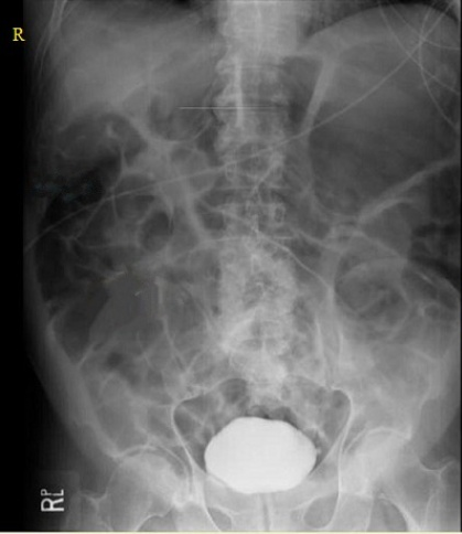
A 56 year old man presents to ED with abdominal discomfort and distention. His bloods are shown below:
- Hb 130 g/L
- Plt 300 x 109/L
- WCC 8 x 10⁹/L
- Na 142 mmol/L
- K 2.8 mmol/L
- Urea 6.5 mmol/L
- Creatinine 145 µmol/L
What is the most likely diagnosis?
Answer:
The x-ray shows generalised distension of small and large intestine with no transition point, and multiple air-fluid levels. This is consistent with a paralytic ileus. Hypokalemia may lead to reduced neural conduction to and within the enteric nervous system, altering the normally highly coordinated reflexes and patterns of GI motility, resulting in reduced or absent peristalsis (paralytic ileus). Other causes of paralytic ileus include: infection e.g. gastroenteritis, electrolyte imbalances e.g. hypomagnesaemia/hyponatraemia, abdominal surgery, endocrine disorders e.g. hypothyroidism, intra-abdominal inflammation/peritonitis, trauma, drugs e.g. opioids, acute mesenteric ischaemia. Patients with ileus typically have vague, mild diffuse abdominal pain, distention, fullness, and bloating. They may report nausea, vomiting, and poor appetite. The abdomen may be distended and tympanic, depending on the degree of abdominal and bowel distention, and may be tender. A distinguishing feature is absent or hypoactive bowel sounds, in contrast to the high-pitched sound of obstruction. The silent abdomen of ileus reveals no discernible peristalsis or succussion splash.Bowel Obstruction
Oncology & Palliative Care / Surgical Emergencies
Last Updated: 9th March 2026
Risk factors
- Previous abdominal surgery with intra-abdominal adhesions (most common cause of small bowel obstruction)
- Intestinal malignancy (most common cause of large bowel obstruction)
- Malrotation
- Crohn's disease
- Hernia with incarceration (Inguinal, ventral incisional, umbilical, and parastomal hernias)
- Appendicitis
- Intussusception
- Volvulus
- Foreign body ingestion
- Diverticular disease
- Radiation enteritis
- Gallstone ileus
- Pelvic mass
Pathophysiology
Small bowel obstruction (SBO) represents an interruption in the patency of the gastrointestinal tract. The proximal dilation of the intestine, together with peristalsis, leads to abdominal cramping (colic), which can become severe. The abdominal pain may also be accompanied by vomiting, while the distal interruption of faecal flow leads to absolute constipation. In acute cases, there can be hyperperistalsis distal to the obstruction, leading to the finding of diarrhoea. Obstructed bowel will, over time, prevent appropriate venous drainage with the possible result of decreased arterial perfusion. Untreated patients develop progressive intestinal ischaemia, necrosis, and perforation.
In large bowel obstruction the colon proximal to the cause of mechanical obstruction dilates and, with increased colonic pressure, mesenteric blood flow is reduced producing mucosal oedema with transudation of fluid and electrolytes into the colonic lumen. This can produce dehydration and electrolyte imbalances. With progression, the arterial blood supply becomes jeopardised with mucosal ulceration, full thickness wall necrosis, and eventual perforation. This provides conditions for bacterial translocation, which can produce septic complications. The caecum is the usual site of rupture, as it has the largest diameter, resulting in faecal soilage of the peritoneal cavity, and sepsis.
Clinical features
Clinical features of bowel obstruction include:
- Symptoms
- Intermittent colicky abdominal pain
- Nausea and vomiting
- Constipation/failure to pass flatus or stool
- Signs of underlying cause e.g. weight loss, rectal bleeding, tenesmus
- Signs
- Abdominal distension
- Abdominal tenderness
- Tympanic abdomen
- High pitched tinkling bowel sounds or absent bowel sounds later
- Signs of peritonitis
- Fever
- Signs of underlying cause e.g. hernia, abdominal mass, rectal mass
Differential diagnosis
- Ileus
- Infectious gastroenteritis
- Toxic megacolon
- Intestinal pseudo-obstruction
- Acute appendicitis
- Acute pancreatitis
Investigations
- Abdominal x-ray
- A plain abdominal radiograph (AXR) is still used in some settings as the initial investigation for bowel obstruction; supine films are initially performed but if not diagnostic may be repeated erect to look for multiple fluid levels in the bowel loops.
- Erect chest x-ray
- May indicate perforation (free subdiaphragmatic air) and the need to consider urgent surgery, but absence of free air does not exclude perforation.
- CT abdomen/pelvis with contrast
- CT is the gold standard investigation for diagnosing bowel obstruction because it has a greater sensitivity than X-ray, can visualise the level and severity of obstruction and is often able to identify the underlying cause.
- CT scans have a high (approximately 90%) accuracy in predicting intestinal strangulation and therefore the need for urgent surgery.
- Bloods
- FBC
- U&Es (dehydration/hypovolaemia, hyponatraemia, hypokalaemia)
- CRP
- Serum lipase/amylase (to exclude pancreatitis)
- Lactate (an elevated lactate reading indicates poor tissue perfusion; it is not diagnostic for intestinal ischaemia)
- ABG (metabolic alkalosis)
- Contrast studies
AXR features:
- Large bowel obstruction
- Peripheral loops of dilated bowel
- Haustra (thicker lines and lines do not cross the full width of the bowel)
- Dilation > 6cm for colon, > 9cm for caecum
- Small bowel obstruction
- Central dilated bowel loops
- Valvulae conniventes (lines are visible across whole width of bowel)
- Dilation > 3cm
Management
- Small bowel obstruction
- Small bowel obstruction is a surgical emergency, with a high risk of morbidity and mortality if not managed correctly.
- Treat in the accident and emergency department with fluid resuscitation, bowel decompression (using a nasogastric tube), and analgesia.
- Use intravenous fluids for patients with signs of shock or severe dehydration, or in patients unable to tolerate oral fluids. In these patients, administer fluid resuscitation and place a catheter to monitor urine output. Correct electrolyte disturbances.
- Pain is one of the predominant symptoms. Assess pain at presentation and throughout the admission.
- Place a nasogastric tube or long intestinal tube to decompress air/fluid in the upper gastrointestinal tract. This may also prevent aerophagia and relieve nausea and vomiting.
- Involve the surgical team early.
- Operative treatment is indicated in patients with complete small bowel obstruction, peritonitis, or evidence of strangulation, and in those who do not respond to non-operative treatment.
- If you suspect ischaemia or strangulation, arrange for surgery as soon as possible, and definitely within 6 hours of the suspected onset of ischaemia or strangulation.
- For patients with adhesional obstruction without signs of peritonitis, strangulation, or bowel ischaemia, try non-operative management for a period of up to 72 hours, but do not delay surgery beyond this point.
- Identify and treat the underlying cause of the bowel obstruction.
- Specific treatment may include appendectomy for appendicitis, tumour resection for obstructing tumour, and hernia repair for inguinal hernia.
- Large bowel obstruction
- Initial management
- All patients, including those with perforation/impending perforation, should be fasted until the underlying cause is resolved.
- Most patients are fluid depleted, so intravenous fluids replace previous losses, and any electrolyte imbalances should be corrected.
- Nasogastric decompression should be part of the initial management of any cause, to decompress the intestinal tract and reduce flow of gastric contents or air towards the obstruction.
- Antibiotics are given preoperatively. Broad-spectrum antibiotics that cover likely pathogens, including amoxicillin, metronidazole, and gentamicin, are recommended.
- Emergency surgery
- Emergency surgery is mandatory in patients with colonic perforation or impending perforation due to obstruction. The objectives of surgical intervention are to deal with intra-abdominal contamination by thorough irrigation, resect the perforated segment, and ideally address the underlying cause.
- Where impending perforation is not suspected, and in the absence of any cause that mandates surgical intervention, a period of conservative management for up to 72 hours may be indicated with treatment of the underlying cause.
- Treatment of underlying cause
- Depends on cause
- Initial management
Complications
- Intestinal perforation
- Intestinal ischaemia +/- necrosis
- Peritonitis
- Sepsis
- Intra-abdominal abscess
- Fluid and electrolyte imbalance
Report A Problem
Is there something wrong with this question? Let us know and we’ll fix it as soon as possible.
Loading Form...
- Biochemistry
- Blood Gases
- Haematology
| Biochemistry | Normal Value |
|---|---|
| Sodium | 135 – 145 mmol/l |
| Potassium | 3.0 – 4.5 mmol/l |
| Urea | 2.5 – 7.5 mmol/l |
| Glucose | 3.5 – 5.0 mmol/l |
| Creatinine | 35 – 135 μmol/l |
| Alanine Aminotransferase (ALT) | 5 – 35 U/l |
| Gamma-glutamyl Transferase (GGT) | < 65 U/l |
| Alkaline Phosphatase (ALP) | 30 – 135 U/l |
| Aspartate Aminotransferase (AST) | < 40 U/l |
| Total Protein | 60 – 80 g/l |
| Albumin | 35 – 50 g/l |
| Globulin | 2.4 – 3.5 g/dl |
| Amylase | < 70 U/l |
| Total Bilirubin | 3 – 17 μmol/l |
| Calcium | 2.1 – 2.5 mmol/l |
| Chloride | 95 – 105 mmol/l |
| Phosphate | 0.8 – 1.4 mmol/l |
| Haematology | Normal Value |
|---|---|
| Haemoglobin | 11.5 – 16.6 g/dl |
| White Blood Cells | 4.0 – 11.0 x 10⁹/l |
| Platelets | 150 – 450 x 109/l |
| MCV | 80 – 96 fl |
| MCHC | 32 – 36 g/dl |
| Neutrophils | 2.0 – 7.5 x 10⁹/l |
| Lymphocytes | 1.5 – 4.0 x 10⁹/l |
| Monocytes | 0.3 – 1.0 x 10⁹/l |
| Eosinophils | 0.1 – 0.5 x 10⁹/l |
| Basophils | < 0.2 x 10⁹/l |
| Reticulocytes | < 2% |
| Haematocrit | 0.35 – 0.49 |
| Red Cell Distribution Width | 11 – 15% |
| Blood Gases | Normal Value |
|---|---|
| pH | 7.35 – 7.45 |
| pO2 | 11 – 14 kPa |
| pCO2 | 4.5 – 6.0 kPa |
| Base Excess | -2 – +2 mmol/l |
| Bicarbonate | 24 – 30 mmol/l |
| Lactate | < 2 mmol/l |