

Trauma
Question 77 of 180
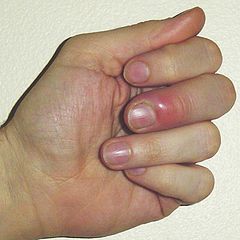
A 30 year old woman presents to the Emergency Department complaining of a red, painful middle finger. What is the diagnosis?
Answer:
An acute paronychia is a localised, superficial infection or abscess of the lateral and proximal skin fold around a nail (perionychium), causing painful swelling. Acute paronychial infections are most often caused by Staphylococcus aureus. Acute paronychia usually affects one finger. Typical features include:- Pain and swelling at the base of the fingernail.
- Localised pain and tenderness of the nail folds.
- Red, tender, and swollen lateral and/or proximal nail folds often with a visible collection of pus.
Hand Injuries
Musculoskeletal / Trauma
Last Updated: 5th September 2022
Clinical signs of hand injury
| Injury | Clinical features |
|---|---|
| Superficial flexor tendon |
|
| Deep flexor tendon |
|
| Complete division of extensor tendon at DIPJ |
|
| Central slip division of extensor tendon at PIPJ |
|
| Median nerve |
|
| Ulnar nerve |
|
| Radial nerve |
|
General principles of treating hand injuries
- Remove rings as soon as possible as swelling can develop relatively rapidly
- Soap or water-based lubricant
- String technique
- Ring-cutters
- Elevate hand to reduce swelling and pain
- Avoid subcutaneous sutures
- X-ray any injury caused by glass
- Consider tetanus prophylaxis
- Assess for tendon injuries and neurovascular status
- Exploration under anaesthetic
- If it is obvious that surgical intervention is required, do not explore the wound in the ED. This particularly applies to suspected nerve injuries where use of LA renders subsequent assessment difficult.
- Conversely, clinical assessment of tendon injuries can be misleading if the patient is reluctant to move due to pain. Exploration under anaesthesia is necessary in this situation and to exclude division of >50% of a tendon (where clinical examination may be normal, but repair is required).
- During exploration, consider the position of the hand at the time of injury— reproducing this may reveal injuries otherwise hidden. Therefore, put all mobile structures through their full range of movement.
Specific types of hand injury
- Extensor tendon injuries
- More than 50% or complete division needs repair by an experienced surgeon.
- Immobilise after repair (e.g. volar slab- type POP with finger joints in full extension and slight flexion at the MCPJs).
- Treat <50% division by splintage in extension (e.g. POP slab as above) under the care of the hand surgeon.
- Flexor tendon injuries
- Refer immediately for specialist repair.
- Nerve injuries
- Complete nerve division may cause surprisingly little sensory loss, so take any altered sensation very seriously. Refer patients with suspected nerve injuries.
- Digital nerves can be repaired up to the level of the DIPJ, although it may be decided not to attempt to repair injuries distal to the PIPJ.
- It is functionally important to have intact sensation over the ‘edges’ of the hand (the thumb, the radial aspect of the index finger, or the ulnar aspect of the little finger).
- Patients sometimes present late after digital nerve injuries— repair can still be quite successful up to 2 weeks after injury.
- Reverse fight bites
- Many human bites occur ‘in reverse’, when an individual punches another in the mouth, causing wounds on the hand over the MCPJs.
- Underlying joint involvement is common and may progress to septic arthritis, unless treated aggressively with exploration, irrigation, and antibiotics. Refer all patients for this.
- Consider hepatitis B, hepatitis C, HIV and tetanus; give appropriate prophylaxis
- Amputations
- Refer patients with partial or complete digital amputation with bone loss.
- Recent proximal amputations without crush injury in fit young patients may be suitable for re- implantation— others may be treated with ‘terminalisation’ or advancement flap.
- Meanwhile, dress, bandage, and elevate; give IV analgesia, tetanus cover, and broad- spectrum antibiotics (e.g. cephalosporin), and keep fasted.
- Wrap the amputated part in moist saline swabs, and place in a sealed plastic bag, surrounded by ice/ water mix at 4°C. Do not freeze or place it directly in solution.
- Finger pad amputations
- Skin loss of <1 cm2 without bony exposure may be allowed to heal with non-adherent dressings.
- Larger areas of tissue loss (particularly in adults) may require skin grafting or advancement flap, but some do heal satisfactorily with simple dressings.
- Ring avulsions
- Refer all circumferential and significant degloving injuries.
- Open (compound) injuries
- Wounds over dislocations or fractures usually require specialist attention.
- Distal open phalangeal fractures may be treated in the ED with wound cleaning, closure, review, and prophylactic antibiotics.
- Crush injuries
- These frequently cause ‘burst’ injury fingertip wounds. Clean the wounds, and take into account the likely swelling when considering closure. Elevate, dress, give analgesia, and arrange review.
- Nail bed lacerations
- Accurate repair may prevent nail deformity.
- Nailfold lacerations extending towards the nail bed require removal of the nail to allow suture.
- Consider replacing the nail after to act as a temporary dressing.
- Foreign bodies under the nail
- Splinters and other FBs under fingernails are relatively common. Apply a digital block and remove with fine forceps. If the FB cannot be reached easily, cut away an appropriate piece of nail.
- Subungual haematomas
- Blood collecting under the nail from a crush injury causes pain.
- If >50% of the nail is affected by a recent injury (within 48hr), trephine the nail distal to the lunula, using a red hot paper clip or battery- operated drill.
Hand fractures and dislocations
- Distal phalangeal fractures
- Treat closed fractures of the distal portion of the distal phalanx with analgesia and elevation.
- Open (compound) burst injuries (from crushing injuries or hammer blows) require meticulous exploration, wound toilet/ repair under LA, and follow- up— local policy will guide if this may be delivered in the ED or under the hand surgeon as an inpatient. Give antibiotics (not a substitute for primary surgical treatment).
- Mallet finger with fracture
- The characteristic ‘mallet finger’ deformity may be associated with a small fracture at the base of the distal phalanx at the point of attachment of the extensor tendon.
- Treat as for (the more usual) mallet finger injury without fracture by plastic mallet splint for 6-7 weeks, advice, and follow- up.
- Refer patients with larger bony fragments (more than one- third of articular surface) with mallet deformity or those with subluxation for possible K- wire internal fixation.
- Proximal and middle phalangeal fractures
- Treat undisplaced fractures with elevation, neighbour strapping, and analgesia.
- Manipulate angulated proximal and middle phalangeal fractures under digital or wrist block.
- A useful tip for proximal phalangeal fractures is to use a needle- holder or a pencil placed adjacent to the web space as a fulcrum.
- Maintain reduction using neighbour strapping and a volar slab POP or flexible padded aluminium (Zimmer) splint, although the latter can be difficult to secure. If reduction is unsatisfactory or cannot be maintained, refer for surgical fixation.
- Index, middle, and ring metacarpal fractures
- Check for displacement or rotational deformity, and refer if either is present.
- Treat with analgesia and elevation, and protect in a volar slab POP.
- Internal fixation may be considered for midshaft MC fractures with marked angulation but can be complicated by marked postoperative stiffness.
- Phalangeal dislocations
- X- ray all dislocations prior to reduction for the presence of associated fractures.
- Reduce under digital or metacarpal nerve block or Entonox® by traction and gentle manipulation, then check the integrity of the collateral ligaments.
- Confirm reduction on X- ray, and immobilise the finger by neighbour strapping. Elevate the hand; provide oral analgesia, and arrange hand clinic follow- up.
- Little (fifth) metacarpal fractures
- These commonly result from punching (Boxer's fracture)
- Check for rotational deformity by gently flexing the fingers into the palm (they should point roughly to the thenar eminence and touch, but not overlap, adjacent fingers on flexion).
- Angulation is common with neck fractures and rarely requires correction, with even up to 40° being accepted.
- Apply neighbour strapping; elevate and give analgesia. Warn the patient that the fifth knuckle will be shorter than before.
- Traditional fracture/ hand clinic follow-up for uncomplicated injuries are increasingly being replaced by written advice with no follow-up (unless problems arise.) Whatever the follow- up arrangements, ensure that the patient is aware of the importance of appropriate hand exercises as soon as possible.
- Refer to the orthopaedic team if there is rotational deformity or significant angulation, particularly with base and shaft fractures, which may need surgery. Also refer patients with associated wounds, remembering that these may be compound human bites.
- Little (fifth) metacarpal dislocations
- Dislocations at the base of the fifth MC may be associated with a fracture.
- Refer for reduction and internal fixation.
Thumb fractures and dislocations
- Dislocation at the metacarpophalangeal joint
- After X- rays and LA block, attempt reduction. If successful, assess and document the integrity of the collateral ligaments, then immobilise in slight (715°) flexion in a POP and arrange follow- up in the fracture clinic. Reduction may be unsuccessful due to ‘button- holing’— in this case, refer for open reduction.
- Gamekeeper’s thumb with associated avulsion fracture
- Most abduction injuries result in ulnar collateral ligament injury without fracture, but occasionally an avulsion fracture occurs at the point of ligament attachment instead.
- Treat this in a scaphoid POP and refer to the fracture clinic, unless the bony fragment is displaced by >2mm, in which case internal fixation will probably be required.
- Thumb dislocations
- Dislocations usually follow falls onto the thumb or hyperextension injuries.
- They can occur at any level, including at the interphalangeal joint (IPJ), MCPJ, and carpometacarpal joint.
- Reduce dislocations by traction and local pressure under combined median and radial nerve blocks.
- Confirm reduction by X- ray; immobilise in a scaphoid POP, and arrange follow- up.
- Bennett’s fracture– dislocation
- This is a fracture through the base of the thumb (first) MC, with radial subluxation of the MC, leaving a small proximal fragment still joined to the trapezium.
- The injury results from a fall onto the thumb or from a fall/ blow onto a fist closed around the thumb.
- Deformity and swelling occur over the base of the thumb and may be mistaken clinically for a scaphoid injury.
- This is an unstable injury requiring expert attention. If undisplaced, apply a Bennett’s- type POP (similar to a scaphoid POP, but with the thumb abducted). If there is any displacement, refer for MUA/fixation. Maintaining reduction often requires the use of screw or Kirschner wire fixation.
Soft tissue hand injuries
- Gamekeeper’s thumb
- The thumb’s ulnar collateral ligament is crucial for stability and function. It is typically injured in hyperabduction injuries (e.g. falls whilst skiing).
- Complete rupture usually results in the two parts of the ligament being separated by the adductor aponeurosis (the ‘Stener lesion’), so satisfactory healing cannot occur.
- If tender over the ulnar collateral ligament of the thumb MCPJ, obtain X- rays— if these demonstrate a fracture, do not stress the joint, but treat appropriately instead.
- If no fracture, assess stability of the ulnar collateral ligament by gentle abduction of the MCPJ (compare with the other hand). Examine the ulnar collateral ligament with the thumb slightly (15°) flexed. If pain precludes adequate examination, consider Entonox® (and/ or LA injection) and repeat the examination.
- Significant (>30°) laxity implies complete rupture and the need for operative repair. Treat uncomplicated sprains with analgesia, elevation, and either crisscross adhesive strapping (‘thumb spica’) or a scaphoid POP if symptoms are severe, and arrange follow- up.
- Mallet finger
- Injury to the extensor mechanism at the DIPJ is relatively common and results from forced flexion of the DIPJ or from a blow/ fall directly onto the fingertip. In the elderly, it can follow minimal trauma.
- There is loss of full active extension at the DIPJ. Normal flexion is preserved.
- X- ray to exclude associated fracture. In the absence of a large fragment, treat in a plastic (mallet) splint secured with tape for ~6wk.
- Ensure the patient understands the need to wear the splint continuously and to keep the finger straight if the splint is removed for washing (e.g. to hold the finger against a flat surface until the splint is replaced). Warn that there may be a small degree of permanent flexion deformity.
- Consider initial follow- up at ~7– 10 days, to ensure compliance with treatment and to reassess in case swelling has decreased and a smaller splint is required.
- Volar plate injury
- These are significant injuries, often with prolonged morbidity. Hyperextension at the PIPJ injures the volar plate at the base of the middle phalanx, with or without evidence of bony involvement.
- Examination shows fusiform swelling of the PIPJ, with tenderness over the volar aspect.
- Treat with ‘buddy strapping’ to adjacent fingers (or ‘Bedford splint’), elevate, provide analgesia, and begin mobilisation immediately. Arrange review to ensure full mobility is regained.
- A2 pulley injury
- The finger flexor tendon sheath at the PIPJ is thickened as the A2 pulley. Occasionally (e.g. in rock climbers), the tendon cuts through the A2 pulley, causing bowstringing on flexion. There may be associated tendon injury.
- Treat with buddy strapping and elevation. Arrange hand specialist follow- up.
- Pulp infections
- Infection of the pulp space at the fingertip may reflect underlying FB or osteomyelitis, so X- ray to search for these and treat accordingly.
- If X- rays are normal, incise the pointing area under LA digital block. Send pus for bacteriology; apply a dressing, commence oral antibiotics (e.g. flucloxacillin 250– 500mg PO qds), and arrange follow- up.
- Paronychia
- Infection of the nailfold adjacent to the nail is common.
- In the early stages, oral antibiotics (e.g. flucloxacillin or clarithromycin) may cure.
- Once pus has developed, drain this under LA digital block by an incision over the fluctuance (usually a small longitudinally orientated incision adjacent to the proximal nailfold suffices, but pus under the nail may require removal of a segment of the nail). Alternatively, incise immediately adjacent to and along the affected lateral nailfold.
- Once drained, do not give antibiotics, unless there is cellulitis or spreading infection or the patient is immunocompromised and/ or has diabetes.
- Pyogenic flexor tenosynovitis
- Infection of a finger flexor tendon sheath may follow a penetrating injury.
- Classically, the evidence is in the form of ‘Kanavel’s signs’:
- Tenderness over the flexor tendon.
- Symmetrical swelling of the finger.
- Finger held in slight flexion.
- Extreme pain on passive extension.
- Consider tetanus prophylaxis and refer urgently for exploration, irrigation, drainage and IV antibiotics.
- Other infections
- These include palmar space infections and septic arthritis— refer immediately for specialist treatment.
- Locked finger
- Elderly patients with underlying osteoarthritis (OA) sometimes present with locking at a finger MCPJ. A fixed flexion deformity is present, such that the patient can flex but not fully extend at the MCPJ. There is usually no particular history of trauma— the underlying cause is entrapment of the palmar plate on an osteophyte.
- Refer for an early hand surgeon opinion— surgery may be required.
- Trigger finger/ thumb
- This is relatively common, but not particularly related to trauma.
- In young children, many resolve spontaneously, although some require surgery. Most cases in adults are satisfactorily treated by steroid injection into the flexor tendon sheath, but leave this to a specialist.
Report A Problem
Is there something wrong with this question? Let us know and we’ll fix it as soon as possible.
Loading Form...
- Biochemistry
- Blood Gases
- Haematology
| Biochemistry | Normal Value |
|---|---|
| Sodium | 135 – 145 mmol/l |
| Potassium | 3.0 – 4.5 mmol/l |
| Urea | 2.5 – 7.5 mmol/l |
| Glucose | 3.5 – 5.0 mmol/l |
| Creatinine | 35 – 135 μmol/l |
| Alanine Aminotransferase (ALT) | 5 – 35 U/l |
| Gamma-glutamyl Transferase (GGT) | < 65 U/l |
| Alkaline Phosphatase (ALP) | 30 – 135 U/l |
| Aspartate Aminotransferase (AST) | < 40 U/l |
| Total Protein | 60 – 80 g/l |
| Albumin | 35 – 50 g/l |
| Globulin | 2.4 – 3.5 g/dl |
| Amylase | < 70 U/l |
| Total Bilirubin | 3 – 17 μmol/l |
| Calcium | 2.1 – 2.5 mmol/l |
| Chloride | 95 – 105 mmol/l |
| Phosphate | 0.8 – 1.4 mmol/l |
| Haematology | Normal Value |
|---|---|
| Haemoglobin | 11.5 – 16.6 g/dl |
| White Blood Cells | 4.0 – 11.0 x 10⁹/l |
| Platelets | 150 – 450 x 109/l |
| MCV | 80 – 96 fl |
| MCHC | 32 – 36 g/dl |
| Neutrophils | 2.0 – 7.5 x 10⁹/l |
| Lymphocytes | 1.5 – 4.0 x 10⁹/l |
| Monocytes | 0.3 – 1.0 x 10⁹/l |
| Eosinophils | 0.1 – 0.5 x 10⁹/l |
| Basophils | < 0.2 x 10⁹/l |
| Reticulocytes | < 2% |
| Haematocrit | 0.35 – 0.49 |
| Red Cell Distribution Width | 11 – 15% |
| Blood Gases | Normal Value |
|---|---|
| pH | 7.35 – 7.45 |
| pO2 | 11 – 14 kPa |
| pCO2 | 4.5 – 6.0 kPa |
| Base Excess | -2 – +2 mmol/l |
| Bicarbonate | 24 – 30 mmol/l |
| Lactate | < 2 mmol/l |