

Trauma
This was previously featured in an exam
This was previously featured in an exam
Question 70 of 216
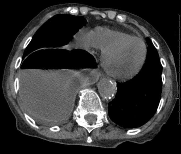
A 60 year old man is brought to ED after losing control of his vehicle whilst intoxicated and hitting a brick wall at approximately 60 mph. His CT chest is shown. What is the diagnosis?
Answer:
The CT shows a right sided diaphragmatic hernia with the stomach in the chest. Note the air fluid level in the stomach. Diaphragmatic injuries are relatively rare and result from either blunt trauma or penetrating trauma. About 80-90% of blunt diaphragmatic ruptures result from motor vehicle collisions (MVCs). Since the pressure is higher in the abdominal cavity than the chest cavity, rupture of the diaphragm is almost always associated with herniation of abdominal organs into the thoracic cavity (diaphragmatic hernia).Thoracic Trauma: Potentially Life-Threatening Injuries
Trauma
Last Updated: 13th February 2021
Unlike immediately life-threatening conditions that are recognised during the primary survey, other potentially lethal injuries are often not obvious on initial physical examination. Diagnosis requires a high index of suspicion and appropriate use of adjunctive studies. The following potentially lethal injuries should be identified and managed during secondary survey:
- Simple pneumothorax
- Haemothorax (massive haemothorax is covered in primary survey)
- Flail chest
- Pulmonary contusion
- Blunt cardiac injury
- Traumatic aortic disruption
- Traumatic diaphragmatic injury
- Blunt oesophageal rupture
Simple pneumothorax
- Pathophysiology:
- Pneumothorax results from air entering the potential space between the visceral and parietal pleura. The thorax is typically completely filled by the lungs, which are held to the chest wall by surface tension between the pleural surfaces. Air in the pleural space disrupts the cohesive forces between the visceral and parietal pleura, allowing the lung to collapse. A ventilation-perfusion defect occurs because the blood that perfuses the non-ventilated area is not oxygenated.
- Both penetrating and non-penetrating trauma can cause this injury. Lung laceration with air leakage is the most common cause of pneumothorax from blunt trauma.
- Diagnosis:
- Perform a comprehensive physical examination of the chest, including inspection for bruising, lacerations, and contusions. Assess movement of the chest wall and assess and compare breath sounds bilaterally.
- When a pneumothorax is present, breath sounds are often decreased on the affected side.
- Percussion may demonstrate hyperresonance, although this finding is extremely difficult to hear in a noisy resuscitation bay.
- An upright expiratory chest x-ray aids in the diagnosis. Patients with blunt polytrauma are not candidates for this evaluation, although patients with penetrating chest trauma may be.
- Management:
- Any pneumothorax is best treated with a chest tube placed in the fifth intercostal space, just anterior to the midaxillary line.
- Observation and aspiration of a small, asymptomatic pneumothorax may be appropriate, but a qualified doctor should make this treatment decision.
- After inserting a chest tube and connecting it to an underwater seal apparatus with or without suction, a chest x-ray examination is done to confirm appropriate placement and reexpansion of the lung.
- Ideally, a patient with a known pneumothorax should not undergo general anaesthesia or receive positive pressure ventilation without having a chest tube inserted. A patient with a pneumothorax should also undergo chest decompression before transport via air ambulance due to the potential risk of expansion of the pneumothorax at altitude, even in a pressurised cabin.
- In selected circumstances, such as when a “subclinical pneumothorax” (i.e. occult) has been diagnosed, the trauma team may decide to carefully observe the patient for signs that the pneumothorax is expanding. The safest approach is to place a chest tube before a tension pneumothorax can develop.
Haemothorax
- Pathophysiology:
- Haemothorax is a type of pleural effusion in which blood (<1500 mL) accumulates in the pleural cavity.
- The primary cause of haemothorax is laceration of the lung, great vessels, an intercostal vessel, or an internal mammary artery from penetrating or blunt trauma.
- Thoracic spine fractures may also be associated with a haemothorax.
- Diagnosis:
- Expose the chest and cervical areas, and observe the movement of the chest wall. Look for any penetrating chest wall injuries, including the posterior thorax. Assess and compare breath sounds in both haemithoraces. Typically, dullness to percussion is heard on the affected side.
- Obtain a chest x-ray with the patient in the supine position. A small amount of blood will be identified as a homogeneous opacity on the affected side.
- Management:
- Bleeding is usually self-limited and does not require operative intervention.
- An acute haemothorax that is large enough to appear on a chest x-ray may be treated with a 28-32 French chest tube. The chest tube evacuates blood, reduces the risk of a clotted haemothorax, and, allows for continuous monitoring of blood loss. Evacuation of blood and fluid also enables clinicians to more completely assess the patient for potential diaphragmatic injury.
- Although many factors are involved in the decision to operate on a patient with a haemothorax, the patient’s physiologic status and the volume of blood drainage from the chest tube are important considerations. Greater than 1500 mL of blood obtained immediately through the chest tube indicates a massive haemothorax that may require operative intervention. Also, if drainage of more than 200 mL/hr for 2 to 4 hours occurs, or if blood transfusion is required, the trauma team should consider operative exploration. The ultimate decision for operative intervention is based on the patient’s haemodynamic status.
Flail chest and pulmonary contusion
- Pathophysiology:
- A flail chest occurs when a segment of the chest wall does not have bony continuity with the rest of the thoracic cage. This condition usually results from trauma associated with multiple rib fractures (i.e. two or more adjacent ribs fractured in two or more places), although it can also occur when there is a costochondral separation of a single rib from the thorax.
- A pulmonary contusion is a bruise of the lung, caused by thoracic trauma. Blood and other fluids accumulate in the lung tissue, interfering with ventilation and potentially leading to hypoxia. Pulmonary contusion can occur without rib fractures or flail chest, particularly in young patients without completely ossified ribs. Children have far more compliant chest walls than adults and may suffer contusions and other internal chest injury without overlying rib fractures. In adults, pulmonary contusion is most often encountered with concomitant rib fractures, and it is the most common potentially lethal chest injury. The resultant respiratory failure can be subtle, developing over time rather than occurring instantaneously. Limited ventilatory reserve may predispose older adult patients to early respiratory failure.
- Diagnosis:
- A flail segment may not be apparent by physical examination, particularly soon after injury. Decreased respiratory effort, combined with contusion and atelectasis, may limit movement of the chest wall. Thick chest wall musculature may also limit visualisation of abnormal chest movement. If the injury results in significant underlying pulmonary contusion, serious hypoxia can result. Restricted chest wall movement associated with pain and underlying lung contusion can lead to respiratory failure.
- Observation of abnormal respiratory motion and palpation of crepitus from rib or cartilage fractures can aid the diagnosis.
- A chest x-ray may suggest multiple rib fractures but may not show costochondral separation.
- Management:
- Initial treatment of flail chest and pulmonary contusion includes administration of humidified oxygen, adequate ventilation, and cautious fluid resuscitation.
- In the absence of systemic hypotension, the administration of crystalloid intravenous solutions should be carefully controlled to prevent volume overload, which can further compromise the patient’s respiratory status.
- Patients with significant hypoxia (i.e. PaO2 < 8.6 kPa or SaO2 < 90%) on room air may require intubation and ventilation within the first hour after injury. Associated medical conditions, such as chronic obstructive pulmonary disease and renal failure, increase the likelihood of requiring early intubation and mechanical ventilation.
- Definitive treatment of flail chest and pulmonary contusion involves ensuring adequate oxygenation, administering fluids judiciously, and providing analgesia to improve ventilation. The plan for definitive management may change with time and patient response, warranting careful monitoring and reevaluation of the patient.
- Analgesia can be achieved with intravenous narcotics or local anaesthetic administration, which avoids the potential respiratory depression common with systemic narcotics. Options for administering local anaesthetics include intermittent intercostal nerve block(s) and transcutaneous intrapleural, extrapleural, or epidural anaesthesia. When used properly, local anaesthetic agents can provide excellent analgesia and prevent the need for intubation.
- Prevention of hypoxia is of paramount importance for trauma patients, and a short period of intubation and ventilation may be necessary until clinicians have diagnosed the entire injury pattern. Careful assessment of the patient’s respiratory rate, arterial oxygen saturation, and work of breathing will indicate appropriate timing for intubation and ventilation, should it be necessary.
Blunt cardiac injury
- Pathophysiology:
- Recent literature review demonstrates 50% of blunt cardiac injury (BCI) was related to motor vehicle crash (MVC), followed by pedestrian struck by vehicles, motorcycle crashes, and then falls from heights greater than 20 feet (6 meters).
- Blunt cardiac injury can result in myocardial muscle contusion, cardiac chamber rupture, coronary artery dissection and/or thrombosis, and valvular disruption.
- Diagnosis:
- Cardiac rupture typically presents with cardiac tamponade and should be recognised during the primary survey. However, occasionally the signs and symptoms of tamponade are slow to develop with an atrial rupture. Early use of FAST can facilitate diagnosis.
- Trauma team members must consider the importance of BCI due to trauma. Patients with blunt myocardial injury may report chest discomfort, but this symptom is often attributed to chest wall contusion or fractures of the sternum and/or ribs.
- The true diagnosis of blunt myocardial injury can be established only by direct inspection of the injured myocardium.
- Clinically significant sequelae are hypotension, dysrhythmias, and/or wall-motion abnormality on two-dimensional echocardiography.
- The electrocardiographic changes are variable and may even indicate frank myocardial infarction. Multiple premature ventricular contractions, unexplained sinus tachycardia, atrial fibrillation, bundle-branch block (usually right), and ST segment changes are the most common ECG findings.
- Elevated central venous pressure with no obvious cause may indicate right ventricular dysfunction secondary to contusion.
- Clinicians must also remember that the traumatic event may have been precipitated by a myocardial ischaemic episode. The presence of cardiac troponins can be diagnostic of myocardial infarction. However, their use in diagnosing blunt cardiac injury is inconclusive and offers no additional information beyond that available from ECG.
- Management:
- Patients with a blunt injury to the heart diagnosed by conduction abnormalities (an abnormal ECG) are at risk for sudden dysrhythmias and should be monitored for the first 24 hours. After this interval, the risk of a dysrhythmia appears to decrease substantially.
- Patients without ECG abnormalities do not require further monitoring.
Traumatic aortic disruption
- Pathophysiology:
- Traumatic aortic rupture is a common cause of sudden death after a vehicle collision or fall from a great height. Survivors of these injuries frequently recover if aortic rupture is promptly identified and treated expeditiously.
- Those patients with the best possibility of surviving tend to have an incomplete laceration near the ligamentum arteriosum of the aorta. Continuity is maintained by an intact adventitial layer or contained mediastinal haematoma, preventing immediate exsanguination and death. Blood may escape into the mediastinum, but one characteristic shared by all survivors is that they have a contained haematoma.
- Persistent or recurrent hypotension is usually due to a separate, unidentified bleeding site. Although free rupture of a transected aorta into the left chest does occur and can cause hypotension, it usually is fatal unless the trauma team can repair it within a few minutes.
- Diagnosis:
- Specific signs and symptoms of traumatic aortic disruption are frequently absent. Maintain a high index of suspicion prompted by a history of decelerating force and its characteristic findings on chest x-ray, and evaluate the patient further. Other radiographic signs of blunt aortic injury include:
- Widened mediastinum
- Obliteration of the aortic knob
- Deviation of the trachea to the right
- Depression of the left mainstem bronchus
- Elevation of the right mainstem bronchus
- Obliteration of the space between the pulmonary artery and the aorta (obscuration of the aortopulmonary window)
- Deviation of the oesophagus (nasogastric tube) to the right
- Widened paratracheal stripe
- Widened paraspinal interfaces
- Presence of a pleural or apical cap
- Left haemothorax
- Fractures of the first or second rib or scapula
- False positive and false negative findings can occur with each x-ray sign, and, infrequently (1%–13%), no mediastinal or initial chest x-ray abnormality is present in patients with great-vessel injury. Even a slight suspicion of aortic injury warrants further evaluation of the patient at a facility capable of repairing the injury.
- Helical contrast-enhanced computed tomography (CT) of the chest has proven to be an accurate screening method for patients with suspected blunt aortic injury. CT scanning should be performed liberally, because the findings on chest x-ray, especially the supine view, are unreliable. If results are equivocal, aortography should be performed. In general, patients who are haemodynamically abnormal should not be placed in a CT scanner. The sensitivity and specificity of helical contrast-enhanced CT have been shown to be close to 100%, but this result is technology dependent. If this test is negative for mediastinal haematoma and aortic rupture, no further diagnostic imaging of the aorta is likely necessary, although the surgical consultant will dictate the need for further imaging.
- Transoesophageal echocardiography (TEE) appears to be a useful, less invasive diagnostic tool. The trauma surgeon caring for the patient is in the best position to determine which, if any, other diagnostic tests are warranted.
- Specific signs and symptoms of traumatic aortic disruption are frequently absent. Maintain a high index of suspicion prompted by a history of decelerating force and its characteristic findings on chest x-ray, and evaluate the patient further. Other radiographic signs of blunt aortic injury include:
- Management:
- Heart rate and blood pressure control can decrease the likelihood of rupture.
- Pain should first be controlled with analgesics.
- If no contraindications exist, heart rate control with a short-acting beta blocker to a goal heart rate of less than 80 beats per minute (BPM) and blood pressure control with a goal mean arterial pressure of 60 to 70 mm Hg is recommended. When beta blockade with esmolol is not sufficient or contraindicated, a calcium channel blocker (nicardipine) can be used; if that fails, nitroglycerin or nitroprusside can be carefully added. Hypotension is an obvious contraindication to these medications.
- A qualified surgeon should treat patients with blunt traumatic aortic injury and assist in the diagnosis. Open repair involves resection and repair of the torn segment or, infrequently, primary repair. Endovascular repair is the most common option for managing aortic injury and has excellent short-term outcomes. Close post-discharge follow-up is necessary to identify long-term complications.
- Low-resourced facilities should not delay transfer by performing extensive evaluations of a wide mediastinum, because free rupture of the contained haematoma and rapid death from exsanguination may occur. All patients with a mechanism of injury and simple chest x-ray findings suggestive of aortic disruption should be transferred to a facility capable of rapid, definitive diagnosis and treatment of this potentially lethal injury.
Traumatic diaphragmatic injury
- Pathophysiology:
- Traumatic diaphragmatic ruptures are more commonly diagnosed on the left side, perhaps because the liver obliterates the defect or protects it on the right side, whereas the appearance of displaced bowel, stomach, and/or nasogastric (NG) tube is more easily detected in the left chest.
- Blunt trauma produces large radial tears that lead to herniation, whereas penetrating trauma produces small perforations that can remain asymptomatic for years.
- Diagnosis:
- Diaphragmatic injuries are frequently missed initially when the chest film is misinterpreted as showing an elevated diaphragm, acute gastric dilation, loculated haemopneumothorax, or subpulmonic haematoma.
- Appearance of an elevated right diaphragm on a chest x-ray may be the only finding of a right-sided injury.
- If a laceration of the left diaphragm is suspected, a gastric tube can be inserted; if the gastric tube appears in the thoracic cavity on the chest film, the need for special contrast studies is eliminated.
- Occasionally, the condition is not identified on the initial x-ray film or subsequent CT scan, in which case an upper gastrointestinal contrast study should be performed.
- The appearance of peritoneal lavage fluid in the chest tube drainage also confirms the diagnosis in patients who have undergone diagnostic peritoneal lavage.
- Minimally invasive endoscopic procedures (e.g. laparoscopy and thoracoscopy) may be helpful in evaluating the diaphragm in indeterminate cases.
- Management:
- Operation for other abdominal injuries often reveals a diaphragmatic tear.
- Treatment is by direct repair.
- Care must be taken when placing a chest tube in patients with suspected diaphragm injury, as tubes can inadvertently injure the abdominal contents that have become displaced into the chest cavity.
Blunt oesophageal rupture
- Pathophysiology:
- Oesophageal trauma most commonly results from penetrating injury.
- Although rare, blunt oesophageal trauma, caused by the forceful expulsion of gastric contents into the oesophagus from a severe blow to the upper abdomen, can be lethal if unrecognised. This forceful ejection produces a linear tear in the lower oesophagus, allowing leakage into the mediastinum.
- The resulting mediastinitis and immediate or delayed rupture into the pleural space causes empyema.
- Diagnosis:
- The clinical picture of patients with blunt oesophageal rupture is identical to that of post-emetic oesophageal rupture.
- The clinical setting of oesophageal injury is typically a patient with a left pneumothorax or haemothorax without a rib fracture who has received a severe blow to the lower sternum or epigastrium and is in pain or shock out of proportion to the apparent injury.
- Particulate matter may drain from the chest tube after the blood begins to clear.
- The presence of mediastinal air also suggests the diagnosis, which often can be confirmed by contrast studies and/or oesophagoscopy.
- Management:
- Treatment of oesophageal rupture consists of wide drainage of the pleural space and mediastinum with direct repair of the injury.
- Repairs performed within a few hours of injury improve the patient’s prognosis.
Report A Problem
Is there something wrong with this question? Let us know and we’ll fix it as soon as possible.
Loading Form...
- Biochemistry
- Blood Gases
- Haematology
| Biochemistry | Normal Value |
|---|---|
| Sodium | 135 – 145 mmol/l |
| Potassium | 3.0 – 4.5 mmol/l |
| Urea | 2.5 – 7.5 mmol/l |
| Glucose | 3.5 – 5.0 mmol/l |
| Creatinine | 35 – 135 μmol/l |
| Alanine Aminotransferase (ALT) | 5 – 35 U/l |
| Gamma-glutamyl Transferase (GGT) | < 65 U/l |
| Alkaline Phosphatase (ALP) | 30 – 135 U/l |
| Aspartate Aminotransferase (AST) | < 40 U/l |
| Total Protein | 60 – 80 g/l |
| Albumin | 35 – 50 g/l |
| Globulin | 2.4 – 3.5 g/dl |
| Amylase | < 70 U/l |
| Total Bilirubin | 3 – 17 μmol/l |
| Calcium | 2.1 – 2.5 mmol/l |
| Chloride | 95 – 105 mmol/l |
| Phosphate | 0.8 – 1.4 mmol/l |
| Haematology | Normal Value |
|---|---|
| Haemoglobin | 11.5 – 16.6 g/dl |
| White Blood Cells | 4.0 – 11.0 x 10⁹/l |
| Platelets | 150 – 450 x 109/l |
| MCV | 80 – 96 fl |
| MCHC | 32 – 36 g/dl |
| Neutrophils | 2.0 – 7.5 x 10⁹/l |
| Lymphocytes | 1.5 – 4.0 x 10⁹/l |
| Monocytes | 0.3 – 1.0 x 10⁹/l |
| Eosinophils | 0.1 – 0.5 x 10⁹/l |
| Basophils | < 0.2 x 10⁹/l |
| Reticulocytes | < 2% |
| Haematocrit | 0.35 – 0.49 |
| Red Cell Distribution Width | 11 – 15% |
| Blood Gases | Normal Value |
|---|---|
| pH | 7.35 – 7.45 |
| pO2 | 11 – 14 kPa |
| pCO2 | 4.5 – 6.0 kPa |
| Base Excess | -2 – +2 mmol/l |
| Bicarbonate | 24 – 30 mmol/l |
| Lactate | < 2 mmol/l |