

Trauma
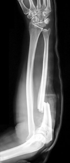
A 34 year old tree surgeon is brought to the Emergency Department by his colleagues after falling from a tree. He is complaining of pain in his right forearm. There is an obvious deformity, a backslab is applied for analgesia and an x-ray performed. What is the diagnosis?
Answer:
Monteggia fracture-dislocation is defined as a fracture of the ulna associated with dislocation of the radial head. It occurs from forced pronation of the forearm (e.g. fall onto an outstretched, fully pronated forearm). It can also occur by a direct blow or fall onto the proximal ulna, displacing the head of the radius. Treat with analgesia and immobilisation in a temporary above- elbow POP backslab. Refer to the orthopaedic team for ORIF (or, sometimes in children, for treatment with MUA and POP).Forearm Injuries
Trauma
Last Updated: 26th January 2023
If one forearm bone is fractured, look for a fracture or dislocation of the other. Obvious deformity in an adult forearm indicates fracture of the radial and ulnar shafts. Initially treat with:
- Analgesia (e.g. increments of IV morphine + antiemetic until pain is relieved).
- Immobilisation in backslab POP.
- If one or both fractures are compound, give IV antibiotics and tetanus cover, and dress the wound.
Always check distal pulses and sensation, and examine for associated injuries at the wrist and elbow. Only once this has been done and the patient is comfortable can he/ she be sent for X- ray. Ensure X- rays demonstrate the whole lengths of the radius and ulna, including separate views of both the elbow and wrist joints.
Fractures of both radial and ulnar shafts
Adult fractures, unlike those in children, may be markedly displaced, with little or no bony contact between the fragments. Rotational deformity is common. Check carefully for clinical evidence of neurovascular injury. Closed reduction is difficult and often fails or is complicated by late slippage. Treat fractures with analgesia/ immobilisation as described earlier, and refer for ORIF.
Isolated ulnar shaft fracture
These usually occur from a direct blow to the outer edge of the forearm (it is typically seen as a defence injury) or from a fall striking the ulnar shaft. X- ray the whole ulna and radius to exclude associated fracture or dislocation of the radial head. If undisplaced, treat in an above- elbow POP, with the elbow flexed to 90° and the forearm in mid- supination. Refer all displaced or angulated fractures for ORIF.
Isolated radial shaft fracture
These are very uncommon. Always treat and assume that there is some associated damage to the distal radioulnar joint at the wrist.
Monteggia fracture-dislocation
This is defined as a fracture of the ulna associated with dislocation of the radial head. It occurs from forced pronation of the forearm (e.g. fall onto an outstretched, fully pronated forearm). It can also occur by a direct blow or fall onto the proximal ulna, displacing the head of the radius. Treat with analgesia and immobilisation in a temporary above- elbow POP backslab. Refer to the orthopaedic team for ORIF (or, sometimes in children, for treatment with MUA and POP).
Note: Monteggia fracture– dislocations are not infrequently missed at initial presentation, due to attention being distracted by the ulna fracture. To avoid this:
- Request elbow and wrist X- rays in any patient with a forearm shaft fracture.
- Check all elbow X- rays carefully to ensure that the radial shaft is normally aligned and the radial head abuts the capitellum: a line (radiocapitellar line) drawn down the shaft of the radius should point to the centre of the capitellum in both AP and lateral views (red line on x-ray below does not point to capitellum)
Galeazzi fracture-dislocation
This is defined as a fracture of the radius associated with dislocation of the distal radioulnar joint at the wrist. Always look for subluxation of the ulna in radial fractures. Treat with analgesia and immobilisation in a temporary POP backslab. Refer for ORIF.
Report A Problem
Is there something wrong with this question? Let us know and we’ll fix it as soon as possible.
Loading Form...
- Biochemistry
- Blood Gases
- Haematology
| Biochemistry | Normal Value |
|---|---|
| Sodium | 135 – 145 mmol/l |
| Potassium | 3.0 – 4.5 mmol/l |
| Urea | 2.5 – 7.5 mmol/l |
| Glucose | 3.5 – 5.0 mmol/l |
| Creatinine | 35 – 135 μmol/l |
| Alanine Aminotransferase (ALT) | 5 – 35 U/l |
| Gamma-glutamyl Transferase (GGT) | < 65 U/l |
| Alkaline Phosphatase (ALP) | 30 – 135 U/l |
| Aspartate Aminotransferase (AST) | < 40 U/l |
| Total Protein | 60 – 80 g/l |
| Albumin | 35 – 50 g/l |
| Globulin | 2.4 – 3.5 g/dl |
| Amylase | < 70 U/l |
| Total Bilirubin | 3 – 17 μmol/l |
| Calcium | 2.1 – 2.5 mmol/l |
| Chloride | 95 – 105 mmol/l |
| Phosphate | 0.8 – 1.4 mmol/l |
| Haematology | Normal Value |
|---|---|
| Haemoglobin | 11.5 – 16.6 g/dl |
| White Blood Cells | 4.0 – 11.0 x 10⁹/l |
| Platelets | 150 – 450 x 109/l |
| MCV | 80 – 96 fl |
| MCHC | 32 – 36 g/dl |
| Neutrophils | 2.0 – 7.5 x 10⁹/l |
| Lymphocytes | 1.5 – 4.0 x 10⁹/l |
| Monocytes | 0.3 – 1.0 x 10⁹/l |
| Eosinophils | 0.1 – 0.5 x 10⁹/l |
| Basophils | < 0.2 x 10⁹/l |
| Reticulocytes | < 2% |
| Haematocrit | 0.35 – 0.49 |
| Red Cell Distribution Width | 11 – 15% |
| Blood Gases | Normal Value |
|---|---|
| pH | 7.35 – 7.45 |
| pO2 | 11 – 14 kPa |
| pCO2 | 4.5 – 6.0 kPa |
| Base Excess | -2 – +2 mmol/l |
| Bicarbonate | 24 – 30 mmol/l |
| Lactate | < 2 mmol/l |