

Ear, Nose & Throat
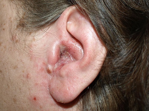
A 62 year old woman presents to the Emergency Department complaining of pain in the left ear. She has no significant past medical history. What is the most likely diagnosis?
Answer:
Clinical features of otitis externa:- Signs:
- The ear canal or external ear, or both, are red, swollen, or eczematous with shedding of the scaly skin.
- Swelling in the ear canal; occasionally this progresses and the swelling eventually completely occludes the ear canal.
- Discharge (serous or purulent) may be present in the ear canal.
- Inflamed eardrum, which may be difficult to visualise if the ear canal is narrowed or filled with debris.
- Symptoms:
- Itch.
- Severe ear pain, disproportionate to the size of the lesion.
- Pain made worse when the tragus or pinna is moved, or when an otoscope is inserted.
- Tenderness on moving the jaw.
- Tender regional lymphadenitis — may be present.
- Loss of hearing if there is sufficient swelling to occlude the ear canal.
Otitis Externa
Ear, Nose & Throat
Last Updated: 12th February 2021
Otitis externa is inflammation of the external ear canal. It may be localised (where there is an infected hair follicle, potentially progressing to a boil in the ear canal) or diffuse where there is more widespread inflammation of the skin and subdermis of the external canal, which can extend to the external ear and the tympanic membrane. It may be acute (< 3 weeks) or chronic (> 3 months). In chronic infection, the lumen of the ear canal progressively narrows and can become completely stenosed over years.
Causes
Acute diffuse otitis externa may be caused by:
- Bacterial infection (most cases) – most commonly Pseudomonas aeruginosa or Staphylococcus aureus
- Fungal infection – most commonly Candida albicans or occasionally Aspergillus species in superficial infection or epidermophyton, trichophyton, and microsporum genera in deeper infections
- Seborrhoeic dermatitis (may have associated dandruff, eyebrow scaling, blepharitis or facial redness/scaling)
- Contact dermatitis – caused by a local irritant or allergen such as topical medications, hearing aids, or earplugs
- Trauma - caused by scratching, aggressive cleaning, ear syringing, foreign objects in the ear, and the use of cotton buds, hearing aids or ear plugs
- Environmental factors — high temperature and/or high humidity, perspiration, swimming (especially in polluted water)
Clinical features
- Signs:
- The ear canal or external ear, or both, are red, swollen, or eczematous with shedding of the scaly skin.
- Swelling in the ear canal; occasionally this progresses and the swelling eventually completely occludes the ear canal.
- Discharge (serous or purulent) may be present in the ear canal.
- Inflamed eardrum, which may be difficult to visualise if the ear canal is narrowed or filled with debris.
- Symptoms:
- Itch.
- Severe ear pain, disproportionate to the size of the lesion.
- Pain made worse when the tragus or pinna is moved, or when an otoscope is inserted.
- Tenderness on moving the jaw.
- Tender regional lymphadenitis — may be present.
- Loss of hearing if there is sufficient swelling to occlude the ear canal.
Diagnosis
Diagnosis is clinical.
Consider taking an ear swab to determine the causative organism if:
- Treatment fails.
- Otitis externa is recurrent or chronic.
- Topical treatment cannot be delivered effectively (for example, if the ear canal is occluded due to swelling or debris).
- The infection has spread beyond the external auditory canal.
- The condition is severe enough to require oral antibiotics.
Management
- Manage any aggravating or precipitating factors.
- Consider cleaning the external auditory ear canal if earwax or debris obstructs the application of topical medication (this may require referral to ENT).
- Syringing or irrigation — to remove debris, provided that the tympanic membrane is intact.
- Dry swabbing — to gently mop out thin secretions from the external auditory canal.
- Microsuction — if irrigation and swabbing are ineffective or inappropriate. This usually requires referral to ENT.
- Prescribe or recommend an analgesic for symptomatic relief, if required.
- Consider prescribing a topical antibiotic with or without a topical corticosteroid.
- Advise the person to use the preparation for a minimum of 7 days, but if symptoms persist to continue using it until they resolve, up to a maximum of 14 days.
- Adverse effects to consider include aminoglycoside-induced ototoxicity in people with a perforated tympanic membrane, aminoglycoside-induced skin sensitization, and fungal superinfection (particularly with long-term use).
- Quinolone containing preparations (for example ciprofloxacin, or ofloxacin) only require twice daily dosing, and can be used in people with a perforated ear drum.
- Topical acetic acid 2% spray is also a safe and effective treatment and can be used for mild cases.
- Consider inserting an ear wick if there is extensive swelling of the auditory canal (this may require ENT referral).
- Oral antibiotics are rarely indicated. Consider seeking specialist advice if an oral antibiotics is thought to be required, including:
- Cellulitis extending beyond the external ear canal.
- When the ear canal is occluded by swelling and debris, and a wick cannot be inserted.
- People with diabetes or compromised immunity, and severe infection or high risk of severe infection, for example with Pseudomonas aeruginosa.
- If an oral antibiotic is to be prescribed in primary care, consider prescribing a 7-day course of flucloxacillin, or clarithromycin (if the person is allergic to penicillin).
- Provide appropriate self-care advice to aid recovery and to reduce risk of future infection:
- Avoid damage to the external ear canal: If earwax is a problem, the person should seek professional advice and have it removed safely to avoid damaging the ear canal. Cotton buds or other objects should not be used to clean the ear canal.
- Keep the ears clean and dry by: Using ear plugs and or a tight fighting cap when swimming — people with acute otitis externa should abstain from water sports for at least 7 to 10 days. Using a hair dryer (at the lowest heat setting) to dry the ear canal after hair washing, bathing, or swimming. Keeping shampoo, soap, and water out of the ear when bathing and showering.
- Ensure skin conditions that are associated with the development of otitis externa are well controlled: If the person is allergic or sensitive to ear plugs, hearing aids, or earrings, they should avoid them, or use alternatives if (for example hypoallergenic hearing aids are available). If the person has a chronic skin condition (for example eczema or psoriasis), they should ensure that this is well controlled if possible.
- Consider using acidifying ear drops or spray (such as EarCalm®) shortly before swimming, after swimming, and at bedtime. These ear drops are available to purchase over the counter at pharmacies.
Complications
The complications of otitis externa include:
- Abscess
- Chronic otitis externa
- Regional dissemination of infection with: auricular cellulitis, chondritis, parotitis, spreading cellulitis
- Fibrosis, leading to stenosis of the ear canal and conductive deafness
- Myringitis (inflammation of the tympanic membrane)
- Tympanic membrane perforation
- Malignant otitis externa
Malignant Otitis Externa
Malignant (necrotising) otitis externa is an extension of otitis externa into the bone surrounding the ear canal. Osteomyelitis will progressively involve the mastoid, temporal and basal skull bones and the infection may spread to the CSF causing meningitis. Pseudomonas aeruginosa is causative in 95% of cases.
Risk factors:
- Diabetes mellitus (most common cause)
- Elderly
- Compromised immunity e.g. HIV/AIDs, chemotherapy or CKD
- Radiotherapy to head and neck
- Aural irrigation with tap water, especially in people with other risk factors
Clinical features:
- Signs:
- Granulation tissue at bone–cartilage junction of ear canal; exposed bone in the ear canal.
- Facial nerve palsy (drooping face on the side of the lesion).
- Temperature over 39°C.
- Lymphadenopathy
- Symptoms:
- Unremitting otalgia, otorrhoea, fever, malaise and headache, more severe than clinical signs would suggest.
- Vertigo.
- Profound hearing loss.
Management:
Patients should be referred urgently to ENT if malignant otitis externa is suspected. Further investigations are carried out in secondary care and include inflammatory markers, radiological imaging, biopsy and culture. Management includes admission, microbiology consultation, treatment with appropriate systemic antibiotics and consideration of surgical treatment.
Report A Problem
Is there something wrong with this question? Let us know and we’ll fix it as soon as possible.
Loading Form...
- Biochemistry
- Blood Gases
- Haematology
| Biochemistry | Normal Value |
|---|---|
| Sodium | 135 – 145 mmol/l |
| Potassium | 3.0 – 4.5 mmol/l |
| Urea | 2.5 – 7.5 mmol/l |
| Glucose | 3.5 – 5.0 mmol/l |
| Creatinine | 35 – 135 μmol/l |
| Alanine Aminotransferase (ALT) | 5 – 35 U/l |
| Gamma-glutamyl Transferase (GGT) | < 65 U/l |
| Alkaline Phosphatase (ALP) | 30 – 135 U/l |
| Aspartate Aminotransferase (AST) | < 40 U/l |
| Total Protein | 60 – 80 g/l |
| Albumin | 35 – 50 g/l |
| Globulin | 2.4 – 3.5 g/dl |
| Amylase | < 70 U/l |
| Total Bilirubin | 3 – 17 μmol/l |
| Calcium | 2.1 – 2.5 mmol/l |
| Chloride | 95 – 105 mmol/l |
| Phosphate | 0.8 – 1.4 mmol/l |
| Haematology | Normal Value |
|---|---|
| Haemoglobin | 11.5 – 16.6 g/dl |
| White Blood Cells | 4.0 – 11.0 x 10⁹/l |
| Platelets | 150 – 450 x 109/l |
| MCV | 80 – 96 fl |
| MCHC | 32 – 36 g/dl |
| Neutrophils | 2.0 – 7.5 x 10⁹/l |
| Lymphocytes | 1.5 – 4.0 x 10⁹/l |
| Monocytes | 0.3 – 1.0 x 10⁹/l |
| Eosinophils | 0.1 – 0.5 x 10⁹/l |
| Basophils | < 0.2 x 10⁹/l |
| Reticulocytes | < 2% |
| Haematocrit | 0.35 – 0.49 |
| Red Cell Distribution Width | 11 – 15% |
| Blood Gases | Normal Value |
|---|---|
| pH | 7.35 – 7.45 |
| pO2 | 11 – 14 kPa |
| pCO2 | 4.5 – 6.0 kPa |
| Base Excess | -2 – +2 mmol/l |
| Bicarbonate | 24 – 30 mmol/l |
| Lactate | < 2 mmol/l |