

Ophthalmology
Question 11 of 97
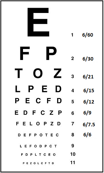
A 56 year old woman presents with a painful, red left eye. On examination you use the Snellen chart shown to assess visual acuity. Using her left eye she is able to read all of line 5 and correctly identifies the letters P and C in line 6. What is the patient's visual acuity in the left eye?
Answer:
- Use a Snellen chart and position patient at a distance of 6m from the chart
- Allow patients to use glasses if available; otherwise use pinhole occluder (made using a needle through a piece of card) which corrects for refractive error
- Ask patient to cover one eye and to read from the top of the chart, until they can no longer read the letters; the smallest line read is their VA
- VA is expressed as: distance from chart in m/distance in m at which a person with normal eyesight is able to read that line of the chart (normal VA is 6/6)
- If patients read additional letters of the line below, record using + number of extra letters (e.g. 6/12 + 2)
- If patients are unable to read the chart at 6m, bring forward metre by metre until they can read the chart, the VA will then be recorded as e.g. 5/60 or 4/60
Acute Red Eye
Ophthalmology
Last Updated: 8th September 2021
Acute red eye is a common presenting complaint.
Differential diagnosis
- Adnexal Causes:
- Trichiasis (posterior misdirection of the eyelashes from the normal site of origin)
- Entropion (inward turning of the eyelid margin)
- Ectropion (outward turning of the eyelid margin)
- Blepharitis (inflammation of the eyelid margin)
- Dry eye (deficiency of the precorneal tear film)
- Conjunctival Causes:
- Conjunctivitis (bacterial, viral, allergic)
- Subconjunctival haemorrhage
- Subtarsal or conjunctival foreign body
- Corneal Causes:
- Infective keratitis (inflammation of the cornea caused by infection)
- Corneal ulcer
- Contact-lens related
- Corneal foreign body
- Corneal abrasion (corneal epithelial defect usually caused by trauma)
- Inflammatory Causes:
- Anterior uveitis (inflammation of the anterior portion of the uveal tract)
- Scleritis (inflammation of the sclera)
- Episcleritis (inflammation of the episclera)
- Traumatic Causes:
- Mechanical
- Chemical injury
- Acute angle-closure glaucoma (closure of the iridocorneal angle leading to an acute rise in intraocular pressure)
Assessment
History:
- Current history
- Acute or gradual onset
- Activity at time of onset
- Duration of symptoms
- Unilateral or bilateral
- Contact lens history
- Associated symptoms
- Visual disturbance
- Pain
- Foreign body sensation
- Discharge
- Itch
- Photophobia
- Eyelid changes
- Past medical and ophthalmological history
- Drug history
Examination:
- Assessment of visual acuity (VA)
- Use a Snellen chart and position patient at a distance of 6m from the chart
- Allow patients to use glasses if available; otherwise use pinhole occluder (made using a needle through a piece of card) which corrects for refractive error
- Ask patient to cover one eye and to read from the top of the chart, until they can no longer read the letters; the smallest line read is their VA
- VA is expressed as: distance from chart in m/distance in m at which a person with normal eyesight is able to read that line of the chart (normal VA is 6/6)
- If patients read additional letters of the line below, record using + number of extra letters (e.g. 6/12 + 2)
- If patients are unable to read the chart at 6m, bring forward metre by metre until they can read the chart, the VA will then be recorded as e.g. 5/60 or 4/60
- If chart cannot be read at 1m, subsequently assess for:
- Counting fingers (record as CF)
- Hand movements (record as HM)
- Light perception (record as PL)
- A hand-held chart at 30 cm is an alternative if a full Snellen chart is not possible
- If patient cannot read letters due to language difficulties, use an E chart - ask patient to state which direction the 3 limbs of the letter point
- Inspection of eyelids
- Look at position of lid margins to exclude trichiasis, entropion or ectropion
- Look for discharge suggestive of conjunctivitis
- Look for swelling/burns suggestive of chemical injury
- Inspection of conjunctiva (including tarsal surface)
- Papillae are see in allergic conjunctivitis
- Follicles are seen in chlamydial conjunctivitis
- Exclude foreign body
- Upper lid should be everted with cotton wool bud to exclude a subtarsal position
- Inspection of ocular surface
- Identify pattern of redness
- Segmental injection may indicate episcleritis or presence of foreign body
- Ciliary or limbal (junction of the cornea and sclera) injection occurs in anterior uveitis and corneal conditions
- Localised and well demarcated redness with quiet surrounding conjunctiva is seen in subconjunctival haemorrhage
- Generalised injection, with engorgement of the deeper scleral vessels and pain on palpation of the globe, indicates the presence of scleritis
- Corneal cloudiness is seen in angle-closure glaucoma
- Perform fluorescein examination
- Allows visualisation of corneal foreign bodies, corneal abrasions and corneal ulcers
- Rose bengal stain can be used in suspected dry eye
- Identify pattern of redness
- Gross inspection of anterior chamber
- Look for blood (hyphema) or pus (hypopyon)
- Assessment of pupillary reactions
- Look for anisocoria (unequal pupil size)
- Look for abnormal pupil shape
- Check for direct and consensual pupillary response (may be abnormal in anterior uveitis or angle-closure glaucoma)
- Assessment of accommodation reflex
- Palpation of globe (If perforation of the globe is suspected (for instance in ocular trauma or as a complication of scleritis), do not palpate the eye)
- Assessment of ocular movements
- Ask about diplopia
- Ask about pain on eye movement
- Look for nystagmus
- Assessment of visual fields
- Fundoscopy
- Slit lamp examination
Red flags
Indications of a serious, and potentially sight-threatening, cause of the person's red eye include:
- Reduced visual acuity
- Deep pain within the eye
- Unilateral red eye
- Contact lens use
- Photophobia
- All high-velocity injuries (for example injuries occurring while hammering or chiseling), or injuries involving glass
- Chemical eye injury
- Ciliary injection
- Fluorescein staining
- Unequal or misshapen pupils, or abnormal pupillary reactions
- Pain on pupillary constriction
- Conjunctivitis in an infant in the first 28 days of life
Refer a person urgently for same-day assessment by an ophthalmologist if they have a suspected serious, and potentially sight-threatening, cause of red eye including:
- Acute glaucoma.
- Corneal ulcer, contact lens-related red eye and corneal foreign body
- Anterior uveitis
- Scleritis
- Trauma, such as penetrating eye injury or high-velocity foreign body
- Chemical injuries.
- Neonatal conjunctivitis
Differential diagnoses causing acute red eye
| Diagnosis | Risk Factors | Symptoms | Signs |
|---|---|---|---|
| Acute angle closure glaucoma | Advancing age, Female gender, Asian ethnicity, Hyperopia, Drugs (adrenergics, antimuscarinics) | Severe eye pain, headache, nausea and vomiting, Visual loss, Lights seen surrounded by halos | Conjunctival injection, Semi-dilated and fixed pupil in oval shape, Hazy oedematous cornea, Tender hard eyeball, Reduced visual acuity, Shallow anterior chamber, Raised intraocular pressure |
| Acute anterior uveitis | Personal history of anterior uveitis, Genetic marker HLA-B27 (associated with ankylosing spondylitis, reactive arthritis, juvenile rheumatoid arthritis) | Eye pain, Blurred vision, Lacrimation, Photophobia | Ciliary flush, Small fixed irregular pupil, Tender eyeball, Positive Talbot's test, Reduced visual acuity, Characteristic slit lamp findings (synechiae, flare, hypopyon, keratic precipitates) |
| Scleritis | Systemic inflammatory or infectious disease (most commonly rheumatoid arthritis) | Severe deep eye pain worse on eye movement, Lacrimation, Photophobia, Visual disturbance | Deep scleral vessel engorgement, Bluish purple sclera discolouration, Tender eyeball, Reduced visual acuity |
| Episcleritis | Usually idiopathic | Eye irritation, Lacrimation | Sectoral redness, Inflammatory nodule |
| Conjunctivitis | Infective: Contact-lens use, Exposure to infected person, URTI, STI; Allergic: Allergen exposure, History of atopy; Irritant: Exposure to environmental irritants | Eye discomfort, Lacrimation, Discharge, Itch | Conjunctival injection, Discharge, Chemosis, Conjunctival follicles or papillae, Eyelid swelling, Preauricular lymphadenopathy |
| Subconjunctival haemorrhage | Usually spontaneous, Hypertension, Bleeding abnormalities, Anticoagulants | Usually asymptomatic | Area of localised well-demarcated haemorrhage |
| Corneal abrasion | Trauma from external objects, Foreign bodies, Contact-lens use | Superficial eye pain, Foreign body sensation, Lacrimation, Photophobia, Blurred vision | Conjunctival injection, Blepharospasm, Fluorescein stains epithelial defect |
| Infective keratitis | Corneal trauma, Contact-lens use, Corneal abrasion/erosion, Foreign body, Previous eye surgery, Dry eyes, Trichiasis | Superficial eye pain, Photophobia, Lacrimation, Discharge, Eyelid oedema, Blurred vision | Reduced visual acuity, Visible corneal ulcer, Cloudy cornea, Corneal vesicles, Fluorescein stains epithelial defect |
Report A Problem
Is there something wrong with this question? Let us know and we’ll fix it as soon as possible.
Loading Form...
- Biochemistry
- Blood Gases
- Haematology
| Biochemistry | Normal Value |
|---|---|
| Sodium | 135 – 145 mmol/l |
| Potassium | 3.0 – 4.5 mmol/l |
| Urea | 2.5 – 7.5 mmol/l |
| Glucose | 3.5 – 5.0 mmol/l |
| Creatinine | 35 – 135 μmol/l |
| Alanine Aminotransferase (ALT) | 5 – 35 U/l |
| Gamma-glutamyl Transferase (GGT) | < 65 U/l |
| Alkaline Phosphatase (ALP) | 30 – 135 U/l |
| Aspartate Aminotransferase (AST) | < 40 U/l |
| Total Protein | 60 – 80 g/l |
| Albumin | 35 – 50 g/l |
| Globulin | 2.4 – 3.5 g/dl |
| Amylase | < 70 U/l |
| Total Bilirubin | 3 – 17 μmol/l |
| Calcium | 2.1 – 2.5 mmol/l |
| Chloride | 95 – 105 mmol/l |
| Phosphate | 0.8 – 1.4 mmol/l |
| Haematology | Normal Value |
|---|---|
| Haemoglobin | 11.5 – 16.6 g/dl |
| White Blood Cells | 4.0 – 11.0 x 10⁹/l |
| Platelets | 150 – 450 x 109/l |
| MCV | 80 – 96 fl |
| MCHC | 32 – 36 g/dl |
| Neutrophils | 2.0 – 7.5 x 10⁹/l |
| Lymphocytes | 1.5 – 4.0 x 10⁹/l |
| Monocytes | 0.3 – 1.0 x 10⁹/l |
| Eosinophils | 0.1 – 0.5 x 10⁹/l |
| Basophils | < 0.2 x 10⁹/l |
| Reticulocytes | < 2% |
| Haematocrit | 0.35 – 0.49 |
| Red Cell Distribution Width | 11 – 15% |
| Blood Gases | Normal Value |
|---|---|
| pH | 7.35 – 7.45 |
| pO2 | 11 – 14 kPa |
| pCO2 | 4.5 – 6.0 kPa |
| Base Excess | -2 – +2 mmol/l |
| Bicarbonate | 24 – 30 mmol/l |
| Lactate | < 2 mmol/l |