

Dermatology
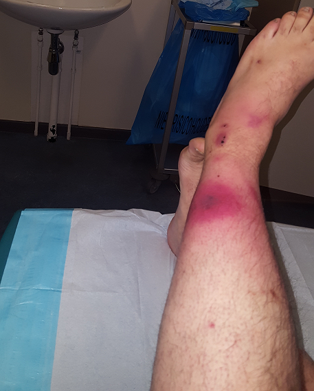
A 54 year old farmer presents to the Emergency Department with a 3 day history of redness and pain over his left lower leg. This has been progressively worsening after the leg was crushed against a fence by a bullock. On examination there is exquisite tenderness in the lower leg with paraesthesia over the area of erythema. His observations recorded at triage are:
- Heart rate: 113 beats/minute
- Blood pressure: 107/69 mmHg
- Respiratory rate: 23 breaths/minute
- Temperature: 37.8°C
What is the diagnosis?
Answer:
Necrotising fasciitis should be suspected in any patient with a soft-tissue infection accompanied by prominent pain and/or anaesthesia over the infected area, or signs and symptoms of systemic toxicity. Altered mental status (Glasgow Coma Scale ≤15), systolic hypotension (systolic BP ≤100 mmHg), and elevated respiratory rate (≥ 22 bpm) suggest that a patient with cellulitis may have a necrotising process requiring expedited surgical evaluation. Examination of the skin overlying the area of cellulitis may reveal crepitus, vesicles, bullae, greyish discoloration, or oedema extending beyond erythema. Subtle skin changes such as leakage of fluid and oedema precede the overt skin changes of blistering and redness. The pain experienced with necrotising fasciitis may be disproportionate to the visible skin changes.Necrotising Fasciitis
Dermatology
Last Updated: 8th September 2021
Necrotising fasciitis is a life-threatening subcutaneous soft-tissue infection that may extend to the deep fascia. About half of cases occur in the extremities, with the remainder concentrated in the perineum, trunk, and head and neck areas.
Causes
The causal organisms may be aerobic, anaerobic, or mixed flora. Two main clinical forms exist.
- Type I necrotising fasciitis is a polymicrobial infection with an anaerobe such as Bacteroides or Peptostreptococcus and a facultative anaerobe such as certain Enterobacterales or non-group A streptococcus. Necrotising fasciitis in the context of recent abdominal surgery or in the groin is most likely to be polymicrobial.
- Type II necrotising fasciitis is most commonly a monomicrobial infection with Streptococcus pyogenes (group A streptococci). The most common site of group A streptococcal necrotising fasciitis is the thigh. Some cases of necrotising fasciitis may have associated myositis due to contiguous spread.
- Other infectious aetiologies may rarely cause a monomicrobial necrotising infection that may be associated with specific exposures or risk factors (e.g. freshwater exposure associated with Aeromonas hydrophila, saltwater exposure or consumption of raw oysters associated with Vibrio vulnificus).
Predisposing risk factors may include diabetes mellitus, peripheral vascular disease, immunocompromising conditions, chronic renal or hepatic insufficiency, chickenpox or herpes zoster, intravenous drug use, or certain medications (e.g. corticosteroids).
Pathophysiology
Bacteria are introduced into the skin and soft tissue from minor trauma, puncture wounds, or surgery. However, in up to 20% of cases no primary site of infection is identified. Infection extends through the fascia but not into the underlying muscle, and tracks along fascial planes extending beyond the area of overlying cellulitis. Systemic signs of necrotising fasciitis, such as fever, tachycardia, and hypotension, are primarily due to the action of bacterial toxins.
Clinical features
Necrotising fasciitis should be suspected in any patient with a soft-tissue infection accompanied by prominent pain and/or anaesthesia over the infected area, or signs and symptoms of systemic toxicity. Altered mental status (Glasgow Coma Scale ≤15), systolic hypotension (systolic BP ≤100 mmHg), and elevated respiratory rate (≥ 22 bpm) suggest that a patient with cellulitis may have a necrotising process requiring expedited surgical evaluation.
Examination of the skin overlying the area of cellulitis may reveal crepitus, vesicles, bullae, greyish discoloration, or oedema extending beyond erythema. Subtle skin changes such as leakage of fluid and oedema precede the overt skin changes of blistering and redness. The pain experienced with necrotising fasciitis may be disproportionate to the visible skin changes.
Differential diagnosis
- Cellulitis
- Erysipelas
- Impetigo
- Myositis
- Gas gangrene
- Cutaneous anthrax
Investigations
No laboratory or imaging studies, alone or in combination, are sufficiently sensitive and specific to definitively diagnose or rule out necrotising fasciitis. All patients admitted with suspected necrotising fasciitis should have a full blood count with white cell differential, urea, electrolytes, creatinine, and C-reactive protein (CRP) measured. Blood cultures should be obtained and may help identify the causative organism. Arterial blood gases may be obtained if there is concern for respiratory compromise.
Necrotising fasciitis is frequently associated with a range of non-specific laboratory abnormalities including:
- Abnormally high or low white blood cell (WBC) count with or without a left shift (elevated percentage of polymorphonuclear leukocytes and/or bands)
- Elevated urea and creatinine due to intracellular volume depletion
- Decreased serum sodium
- Elevated CRP
- Elevated serum creatine kinase
- Elevated plasma lactate
- Decreased serum bicarbonate
Imaging studies should not delay surgical intervention when diagnosis is suspected. In clinically stable patients, imaging may provide supportive evidence for a necrotising process. Plain radiography is frequently normal during the early stages; subcutaneous gas may be present as the disease progresses. The diagnosis should be strongly suspected if soft-tissue gas is visualised on radiological examination, which may also demonstrate abnormalities in the involved soft tissue. Computed tomography and magnetic resonance imaging offer higher sensitivity.
Definitive bacteriological diagnosis is best made from tissue specimens obtained from surgical debridement. Staining of clinically affected tissue may provide an early indication of the causative organism(s).
Management
An urgent surgical consultation should be obtained as soon as the diagnosis is suspected. Treatment should not be delayed while awaiting microbiological and imaging investigations.
Definitive treatment is surgical debridement, repeated as necessary. When debridement is performed, surgical incisions should extend beyond the areas of visible necrosis and the entire necrotic area excised. Surgical specimens including tissue and fluid should be obtained for microbiological culture.
Antibiotic therapy is crucial, but is considered adjunctive to surgical management. Empirical antibiotics should cover major bacterial aetiological agents, and group A streptococcal toxin production that can accompany type II necrotising fasciitis. Tailoring of antimicrobial therapy, as appropriate, is recommended when causative microbial organism(s) are identified via culture.
Report A Problem
Is there something wrong with this question? Let us know and we’ll fix it as soon as possible.
Loading Form...
- Biochemistry
- Blood Gases
- Haematology
| Biochemistry | Normal Value |
|---|---|
| Sodium | 135 – 145 mmol/l |
| Potassium | 3.0 – 4.5 mmol/l |
| Urea | 2.5 – 7.5 mmol/l |
| Glucose | 3.5 – 5.0 mmol/l |
| Creatinine | 35 – 135 μmol/l |
| Alanine Aminotransferase (ALT) | 5 – 35 U/l |
| Gamma-glutamyl Transferase (GGT) | < 65 U/l |
| Alkaline Phosphatase (ALP) | 30 – 135 U/l |
| Aspartate Aminotransferase (AST) | < 40 U/l |
| Total Protein | 60 – 80 g/l |
| Albumin | 35 – 50 g/l |
| Globulin | 2.4 – 3.5 g/dl |
| Amylase | < 70 U/l |
| Total Bilirubin | 3 – 17 μmol/l |
| Calcium | 2.1 – 2.5 mmol/l |
| Chloride | 95 – 105 mmol/l |
| Phosphate | 0.8 – 1.4 mmol/l |
| Haematology | Normal Value |
|---|---|
| Haemoglobin | 11.5 – 16.6 g/dl |
| White Blood Cells | 4.0 – 11.0 x 10⁹/l |
| Platelets | 150 – 450 x 109/l |
| MCV | 80 – 96 fl |
| MCHC | 32 – 36 g/dl |
| Neutrophils | 2.0 – 7.5 x 10⁹/l |
| Lymphocytes | 1.5 – 4.0 x 10⁹/l |
| Monocytes | 0.3 – 1.0 x 10⁹/l |
| Eosinophils | 0.1 – 0.5 x 10⁹/l |
| Basophils | < 0.2 x 10⁹/l |
| Reticulocytes | < 2% |
| Haematocrit | 0.35 – 0.49 |
| Red Cell Distribution Width | 11 – 15% |
| Blood Gases | Normal Value |
|---|---|
| pH | 7.35 – 7.45 |
| pO2 | 11 – 14 kPa |
| pCO2 | 4.5 – 6.0 kPa |
| Base Excess | -2 – +2 mmol/l |
| Bicarbonate | 24 – 30 mmol/l |
| Lactate | < 2 mmol/l |